
SKU : SCG-W5000 PLUS
หมวดหมู่ : Gel Documentation , 12. Lab Equipment , Laboratory Instrument , Servicebio ,
แบรนด์ : Servicebio
Share
Multi-functional Imaging System
Integrated with a chemiluminescence system and a gel imaging system, it can be used for experiments such as Western Blotting membrane detection, nucleic acid gel imaging/gel cutting, and protein gel imaging.
Product Information
The SCG-W5000 PLUS is a comprehensive device that integrates chemiluminescence technology and gel imaging. it is equipped with a high-sensitivity cooled camera with 9 million pixels, enabling rapid, accurate, and high throughput detection and imaging of samples. it is widely used in the fields of life sciences, medicine, and environmental protection.
SOFTWARE USER MANUAL:
SWE Imaging Grayscale Analysis Software-SCG-W2000 SCG-W3000.pdf
Product Features
Chemiluminescence Imaging System :
Obtain Western blot results in a single imaging session, regardless of signal strength—weak signals can be further enhanced by extending the imaging time directly, while strong signals allow retrospective adjustment to any captured image within the exposure timeline.
Supports simultaneous imaging of up to 10 membranes or more, significantly improving instrument efficiency—one system can perform the work of 10 conventional instruments.
Enables color Marker imaging with high sensitivity, delivering exceptional performance even for weak signals.
Marker optimization function eliminates non-specific binding signals between pre- stained markers and antibodies, while also allowing adjustment of overexposed or underexposed luminescent marker signals.
Source files are saved for flexible reprocessing—bands that appear too thick or too faint can be optimally adjusted on a computer post-experiment.
Protein gel imaging capability: captures both color and monochrome protein gel images, with color results providing more intuitive visualization.
Nucleic acid gel imaging: equipped with standard 310/254/365 nm LED UV light sources, compatible with common nucleic acid dyes such as EB, SerRed, SerBlue, GoldView, and SYBR Green. LED UV ensures uniform illumination, superior image quality, and longer lifespan.
Nucleic acid gel excision: includes a standard UV laser protection plate with a cutoff depth > OD7, offering high protection levels for safer gel cutting!
Gel Imaging System:
Three modes and multiple parameters can be flexibly adjusted, resulting in clear and bright imaging
UV laser protective board, safe and convenient for gel cutting
TechnicalSpecifications
| Product Name | Multi-functional Imaging System | |
| Cat.No. | SCG-W5000 PLUS | |
| Dimensions | 400×371×700 mm | |
| Camera | PixelResolution | 9 million |
| Resolution | 2992×3000 | |
| Pixel size | 3.76×3.76μm | |
| Target size | 1(11.28×11.28 mm) | |
| Full Well Capacity | 16.5ke-(HCG),50.5ke-(LCG) | |
| Sensitivity | 877mv@1/30s | |
| ReadoutNoise | 1.24e-(HCG),3.22e-(LCG) | |
| Dark Current | 0.0003e-/s/pixel@-15 | |
| Signal-to-Noise Ratio | 42.2dB(HCG),47dB(LCG) | |
| ExposureTime | 0.1ms~1h | |
| Binning Mode | 1×1,2×2,3×3 | |
| Grayscale | 16-bit(65536 levels) | |
| Cooling | Relative to Ambient Temperature -40°c | |
| Camera Type | Black and White Camera | |
| Lens | Aperture | F0.95-F16 |
| Focal Length | 17mm | |
| Type | Motorized zoom lens | |
Light Source | Bright Field Light source | Downward-facing LED white light source, symmetrically distributed on both sides |
| Ultraviolet light source | 310nm LED array with uniform transmittedillumination,254nm/365nm LED ultraviolet light sources (symmetrically distributed on both sides) | |
| Dark Box | Light isolation | Fully light-sealed, isolates environmental light. |
| Door Control | The door control sensor can control the on/off of the bright field light source. | |
| Rotating disc | Switch the filter according to the current mode to match the applications of chemiluminescence and gelimaging. | |
| Field of View | Effective field of view for membrane imaging is 140mmx 140mm Effective field of view for protein gel imagingis140mmx 140mm The effective field of view for nucleic acid gel imaging is 140mmx140mm | |
| Gel Cutting | After opening the door, the UV light source can be extracted and used with aUV protective board for cutting adhesive | |
Software Functions | Exposure Modes | High Quality: Image quality is the highest |
| Auto Exposure | Intelligent exposure technology quickly determines the optimal exposure time. With the combination of time imaging and time accumulation functions, users can achieve the best image results with just one operation. | |
| Real-time imaging | Real-time presentation of the changes in sample signals during the exposure process, allowing for the observation of every detail of the capture. Overexposed areas will be indicated for samples with overexposure. | |
| Time imaging | After exposure is complete, each frame image within the exposure time can be generated Through precise retrospective adjustments, users can choose any frame image within that exposure time as the final output. | |
| Time Accumulation | For samples with insufficient exposure, users can choose to continue exposure after the initial exposure is completed, enabling the sample to receive additional exposure on top of the already exposed time. | |
| IndustrialComputer | 10.4"display(1024x768)Windows 10 0s 16GB RAM,512GB SSD,Integrated Bluetooth/Wi-Fi | |
| ExternalInterfaces | USB 3.0×2 | |
| Operating Voltage | 90~132VAC/180~264VAC(selectable viaswitch),47~63Hz | |
| ProductPower | 300W | |
| Product Net Weight | 30.65Kg | |
Notes
It is prohibited to touch or scratch the internal lenses of the dark box with hands or sharp objects
After placing the experimental samples, make sure to close the instrument's flip door to prevent external light from entering the dark box and affecting the experimental results
During imaging experiments, shaking the experimental table or instrument is prohibited to avoid impacting the image quality. Pay attention to electrical safety. Pullingor moving the power cord during the experiment is prohibited After the experiment is completed, clean the samples and any residues inside the dark box thoroughly
| Cat. No. | SCG-W5000 | SCG-W3000 | SCG-W1000 |
| Dimension | 400×371×700 mm | 400×371×700 mm | 400×371×700 mm |
| Camera | Depth-cooled high sensitivity camera | Depth-cooled high sensitivity camera | High-sensitivity camera |
| Resolution | 2992*3000,9 megapixels | 2992*3000, 9 megapixels | 3072*2048,4.2 megapixels |
| Pixel | 3.76×3.76 μm | 3.76×3.76 μm | 2.4×2.4 μm |
| Shooting Area | Effective field of view for blotting film/protein gel: 140 x 140 mm Effective field of view for nucleic acid gel: 140 x 140 mm | Blotting Film 136×136 mm | Nucleic Acid Gel / Protein Gel 240×240 mm |
| Cooling Temperature | Relative ambient temperature -40°C | Relative ambient temperature -40°C | - |
| Light Source | Brightfield light source: Epi-illumination LED white light source, symmetrically distributed on both sides UV light source: 310 nm LED array for uniform transmission illumination; epi-illumination 254 nm/365 nm LED UV light source, symmetrically distributed on both sides Blue/white dual light source (optional accessory): Blue/white transmission switching, with 3-level power cycling adjustment for each. | Downward-facing LED white light, symmetrically distributed on both sides | Brightfield illumination: Epi-illumination LED white light source, symmetrically distributed on both sides UV illumination: 310 nm LED array for uniform transmission illumination Blue/white dual light source (optional): Blue/white transmission switching with 3-level adjustable power for each mode |
| Industrial Computer | 10.4 inches, 1024×768 Windows operating system | 10.4 inches, 1024×768 Windows operating system | 10.4 inches, 1024×768 Windows operating system |
| External Interface | 2 USB3.0 | 2 USB3.0 | 2 USB3.0 |
| Working Voltage | 90~132VAC/180~264VAC(selectable viaswitch),47~63Hz | 90~132VAC/180~264VAC | 90~132VAC/180~264VAC(selectable viaswitch),47~63Hz |
| Product Power | 300W | 200W | 300W |
| Net Weight | 30.65 kg | 23.45 kg | 30.65 kg |
| Real-Time Imaging | Yes | Yes | - |
| Time Imaging | Yes | Yes | - |
| Time Accumulation | Yes | Yes | - |
| Auto Exposure | Yes | Yes | Yes |
| Choice of 3 Imaging Modes | Yes | Yes | - |
| Protein Gel/Nucleic Acid Gel Imaging | Yes | - | Yes |
| Nucleic Acid Gel Cutting | Yes | - | Yes |
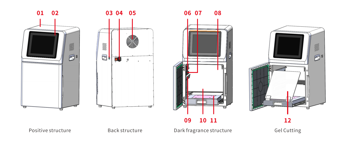
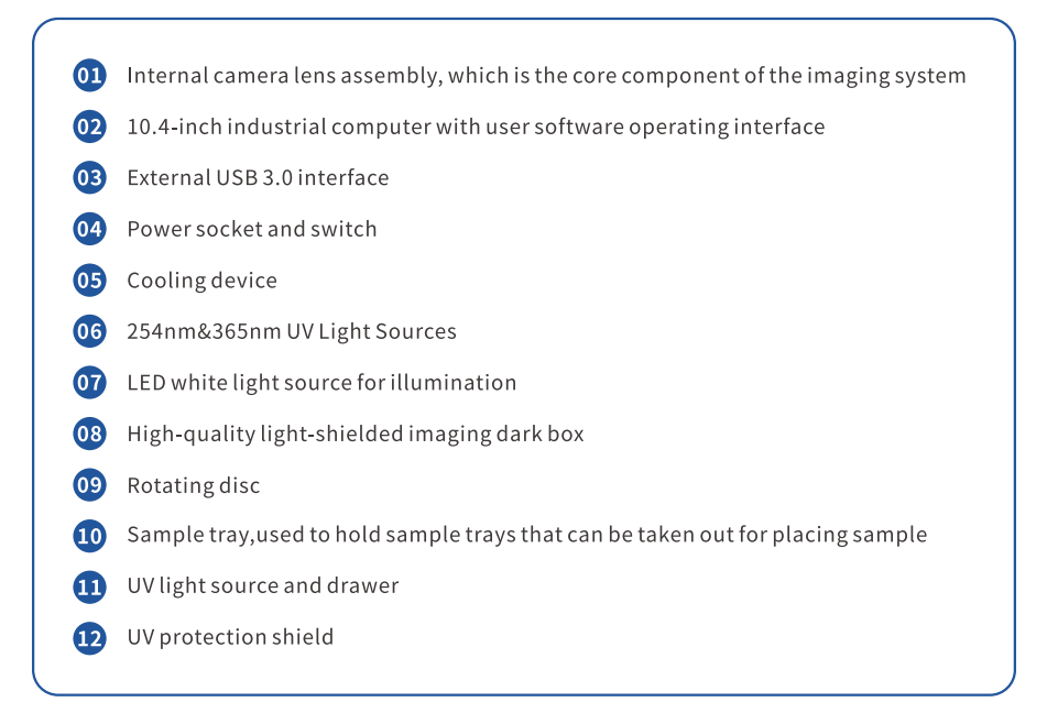
Functional Description


