
SKU :
Option name 1
Categories : Biochemicals , 1. Chemical and Reagents , Buffers , Servicebio ,
Brand : Servicebio
Share
Product Information
Product Name | Cat.No. | Spec. |
Solid Blue Dye | G1044-100ML | 100mL |
G1044-500ML | 500mL |
Product Description/Introduction
This product can be used to dye the myelin sheath. The myeLin sheath is composed of sphingomyelin, which are mainly lipid-like and proteins. It is a segmented tubular sheath wrapped around the axon of the nerve, which acts as insulation and increases the conduction speed of nerve impulses, and plays a role in protecting axons in the central and peripheral nervous system. LuxoL fast bLue has the property of binding to myelin and coloring the myelin sheath.
Storage and Shipping Conditions
Ship and store at room temperature, valid for 18 months.
Assay Protocol/Procedures
1. Dewaxing paraffin sections to water.
2. Preheat the solid blue dye solution in the oven at 65 for 30min, stain slice with the preheated solid blue dye solution at 65 for 4h (covered).
3. Take the dyeing kit out of the oven and cool it for 5-10min, then take out the slices and wash them with tap water until the slide is colorless.
4. The sections are immersed in lithium carbonate and differentiated slightly for 2s (while still hot), wash with water to terminate the differentiation, examine under microscope, and differentiated repeatedly with water and microscope until the myelin sheath is blue with nearly colorless background. If the background is not easy to differentiate and fade, the sections are alternately immersed in lithium carbonate and ethanol for differentiation (alternating between the two solutions without washing), wash to terminate the differentiation, microscopic examination, and repeatedly alternate differentiated washing and microscopic examination until the myelin is blue and the background is nearly colorless.
5. Cut the slices into three tanks of anhydrous ethanol for dehydration, 5min each, xylene transparent for 5min, and neutral gum seal the slices. The slices were dehydrated in 3 cylinders of anhydrous ethanol for 5 min each, transparent in xylene for 5 min and sealed with neutral gum.
6. Microscopic staining results: the myelin sheath of the nerve is blue, the background is light blue or almost colorless.
Note
1. The solid blue dyeing solution needs to be preheated before dyeing, and the temperature of the dyeing solution will have a certain influence on the strength of tissue coloring.
2. After the section is washed, it needs to be differentiated as soon as possible, and the color of the tissue will fade when placed in water for a long time; Prolonged exposure to air will dry out the tissue, resulting in uneven color during differentiation.
3. If you need a kit, you can buy the G1030 myelin dye kit.
4. Each 100 mL of Staining Solution can be used to stain (dip) approximately 60 sections. Replace the stain with a new one when the tissue or cell coloring is significantly lighter or abnormal.

Spinal cord: The gray matter is colorless or light blue. Blue filaments can be seen in the gray matter.
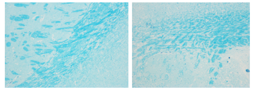
Brain: Blue myelin sheath, light blue or nearly colorless elsewhere.
Nerves: The myelin sheath is blue, mostly colorless elsewhere.
For Research Use Only!

")
")
, 10 plates (for Molecular cloning medium)")
")
")
, 10 plates (for Molecular cloning medium)")